Explainer · July 2, 2026 · 5 min · By Renata Oyola
The Stretch Test: How Clinicians Tell Pigment, Shadow, and Vascular Dark Circles Apart
Most under-eye treatments fail because they target the wrong cause. A two-minute bedside exam, used by dermatologists for decades, can tell you which of the three main types you actually have.
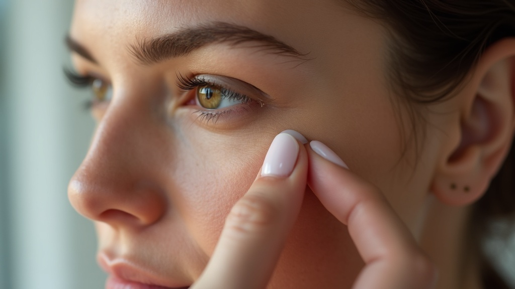
Walk into any pharmacy and the eye cream aisle treats dark circles as a single problem. Clinically, they are at least three distinct problems that happen to share a location. Pigmented circles are a melanin issue. Vascular circles are a blood flow and skin thickness issue. Structural circles are not a discoloration at all, they are a shadow cast by the shape of the face. Each responds to a different intervention, and each is made worse by the wrong one. That is why dermatologists often begin an under-eye consultation with a maneuver so simple it sounds like folk medicine: they stretch the skin.
How the stretch test works
The patient looks straight ahead in even, diffuse light. The clinician places a fingertip just below the darkened area and gently pulls the skin downward and slightly outward, flattening the under-eye contour. Then they watch what the darkness does.
If the discoloration stays exactly where it is and moves with the skin, the color lives in the skin itself. That points to pigmentation: excess melanin deposited in the epidermis or upper dermis. This pattern is common in people with medium to deep skin tones, often runs in families, and is frequently worsened by rubbing the eyes, chronic allergic inflammation, or sun exposure. Some clinicians add a Wood's lamp, a handheld ultraviolet light, to refine the picture. Epidermal melanin tends to appear more sharply defined under the lamp, while deeper dermal pigment looks blurred, which matters because superficial pigment responds better to topical agents than deep pigment does.
If the darkness improves or disappears when the skin is stretched, the cause is usually structural. The circle was never a color problem. It was a shadow created by a tear trough depression, mild under-eye hollowing, or a small fat pad above the trough that catches overhead light. Stretching temporarily smooths the contour, so the shadow vanishes. A quick confirmation: photograph the area with light coming from directly in front of the face rather than above. Shadow-type circles fade dramatically in frontal light. Pigment-type circles do not care where the light comes from.
If the area looks bluish or purplish and blanches briefly when pressed, the culprit is vascular. The skin below the eye is among the thinnest on the body, often under one millimeter, and in some people it is thin enough that the network of small veins and capillaries beneath it shows through. Fatigue, dehydration, nasal congestion, and alcohol can all dilate these vessels or slow drainage, which is why vascular circles fluctuate day to day while pigmented circles are stubbornly constant.
Why the distinction changes everything
Consider what happens when the types are mismatched with treatment. A brightening cream built around vitamin C, azelaic acid, or kojic acid works by interrupting melanin production. Applied to a structural shadow, it does nothing, because there is no excess melanin to suppress. Applied to a vascular circle, it also does nothing, because the color is hemoglobin seen through thin skin, not pigment in the skin.
Caffeine-based products, which constrict superficial vessels temporarily, can modestly help vascular circles for a few hours but will not touch pigmentation or shadow. Retinoids occupy an interesting middle ground: over months, they can thicken the dermis slightly, which makes thin-skin vascular circles less visible, and they accelerate turnover of pigmented cells. But retinoids can also irritate the delicate periorbital skin, and irritation itself can trigger post-inflammatory pigmentation in susceptible skin tones, converting one type of circle into two.
Structural circles generally require a contour-based approach, which is why they are the type most often discussed in the context of injectable fillers or, less invasively, careful light management: makeup artists have long known that a slightly lighter, peach-toned concealer placed only in the trough works by filling the shadow optically rather than covering a color.
The mixed picture problem
Here is the complication clinicians emphasize: most adults over thirty have a combination. A common pattern is mild tear trough hollowing plus thin skin plus a little allergy-driven pigment from years of eye rubbing. In mixed cases the stretch test still helps, because it estimates the proportions. If stretching removes eighty percent of the darkness, contouring approaches will do most of the work. If it removes almost none, pigment or vascular strategies deserve priority.
What you can do at home
You can run a rough version of this exam yourself with a mirror, indirect daylight, and a gentle fingertip pull. Note whether the color moves, fades, or blanches. Photograph the area in overhead light and again in frontal light. None of this replaces an in-person evaluation, especially if darkening is new, one-sided, or accompanied by swelling, which warrants medical assessment. But it will make you a far more informed reader of ingredient labels, and it explains a frustration many people share: the product was not necessarily bad. It was simply answering a question your under-eyes never asked.
Related reading: Allergies: the most overlooked cause of dark circles.